


【3D Printing】Bambu Lab H2D Aids Veterinary Orthopedic Surgery
How 3D Printing Technology Helps Veterinary Surgeons Rehearse Procedures
How 3D Printing Technology Helps Veterinary Surgeons Rehearse Procedures
When performing surgery on patients who cannot speak, veterinary surgeons experience a level of stress and anxiety that is hard for others to imagine. Human surgeons can understand symptoms through patient descriptions, confirm pain locations, and obtain consent after fully understanding the procedure. However, veterinary surgeons do not have these conveniences. They must rely entirely on scan images, extensive experience, and the mental ability to convert two-dimensional images into three-dimensional problems, which they then solve with their hands.
In a surgical field with an extremely low tolerance for error, this mental transformation process is precisely where mistakes can occur.
Orthopedic surgery presents even greater challenges. Bones are irreversible; if a screw is misplaced, the cutting angle is incorrect, or the brace does not perfectly match the anatomy, these are not trivial issues. They can lead to secondary surgeries, prolonged recovery periods, or even worse outcomes. In veterinary orthopedics, anatomical variability is immense: the femurs of a Great Dane and a Dachshund differ not only in size but also significantly in structure, posing severe challenges for standardized implants and surgical methods.
Traditionally, the solution to this problem has relied on experience. Through continuous surgery, a knowledge base of anatomical variations is accumulated, and veterinarians learn to anticipate situations before they occur.
This approach is effective to a certain extent.
However, its limitations are that experience is personal, non-transferable, and always struggles to cope with the next unusual case.
For decades, the medical community has been quietly developing another solution: pre-operative rehearsal. The concept is straightforward—if practice can be performed before actual surgery, the uncertainty of the procedure can be greatly reduced. Pilots practice in simulators, and architects create scale models. Surgeons, too, are increasingly utilizing 3D-printed anatomical models for pre-operative rehearsal.
A physical model that precisely represents the bone to be operated on—not a textbook illustration or a rendering on a screen, but an object that can be held, rotated, and physically manipulated—fundamentally changes the nature of surgical preparation. Surgeons no longer just imagine the surgical process; they actually rehearse it. They can discover problems before intraoperative incidents occur, test implant fit, plan screw trajectories, and confirm the surgical plan before the patient is anesthetized.
However, translating this logic into reliable clinical practice is far more complex than it sounds. This technology requires sufficient precision to be anatomically meaningful; it needs to be fast enough to integrate into actual clinical workflows; and it must be able to produce truly usable models, not approximations full of internal defects or composed of misaligned fragments.
For a long time, meeting these requirements simultaneously has been a formidable challenge.
| Dr. Sebastian's Challenge and Bambu Lab's Solution
Dr. Sebastian has a deeply personal understanding of this problem and has dedicated significant effort to finding a solution. Today, with the help of Bambu Lab 3D printing technology, he can perform precise pre-operative rehearsals before surgery.
Dr. Sebastian is a veterinary doctor specializing in complex surgical procedures. To better prepare for surgery and reduce patient anesthesia time, he has developed a rigorous pre-operative workflow: scanning injured bone structures, modeling surgical steps in software, then 3D printing physical replicas, and rehearsing the surgery directly on them.
By routinely converting medical scans into physical bone models, he can practice in advance, thus entering the operating room with greater precision and confidence.
| Challenges: Precision, Efficiency, and Safety
Although 3D printing has become central to Dr. Sebastian's workflow, the technologies he previously relied on had practical limitations. Resin printers did offer good precision, but hollow bone models required internal support structures that would be permanently trapped within the medullary cavity.
As a result, these models were difficult to use during surgical rehearsal and were compromised in the most critical anatomical areas.
Resin-printed bone model, with linear support structures clearly visible at the bone joints.
Larger or more complex bones had to be printed in segments and then assembled, which increased preparation time and introduced inaccuracies.
Furthermore, the resin printing process itself introduced additional burdens: toxic substances produced during post-processing meant Dr. Sebastian had to maintain a dedicated air handling system—a significant complexity for a tool intended to simplify his work.
| Bambu Lab's Solution
Bambu Lab sponsored Dr. Sebastian with a H2D dual-extruder large-format 3D printer. The dual-extruder system supports printing with soluble support materials, and the spacious build volume allows for the one-time production of full-size bone models, whenever the geometry allows.
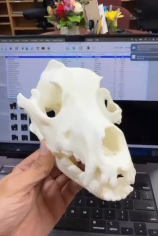
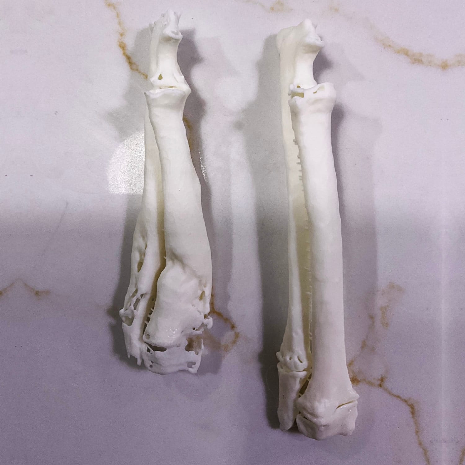
Skull model printed with soluble support material; as shown, the support material has been completely removed.
Soluble support materials have revolutionized how internal structures are printed. After printing, the support material completely dissolves, leaving a clean medullary cavity that closely matches the true anatomy—no residue, no compromise.
This directly addresses the most significant limitation Dr. Sebastian faced when using resin printing.
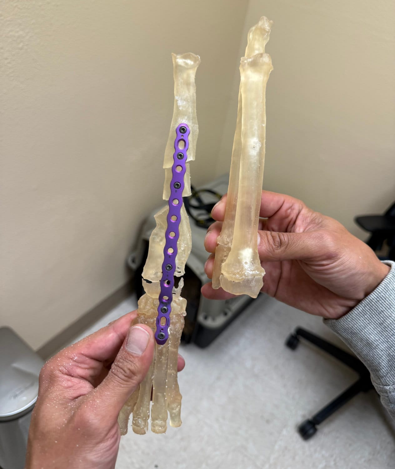
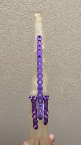
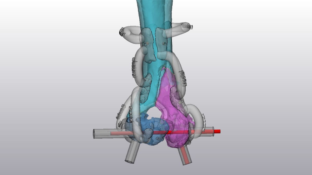
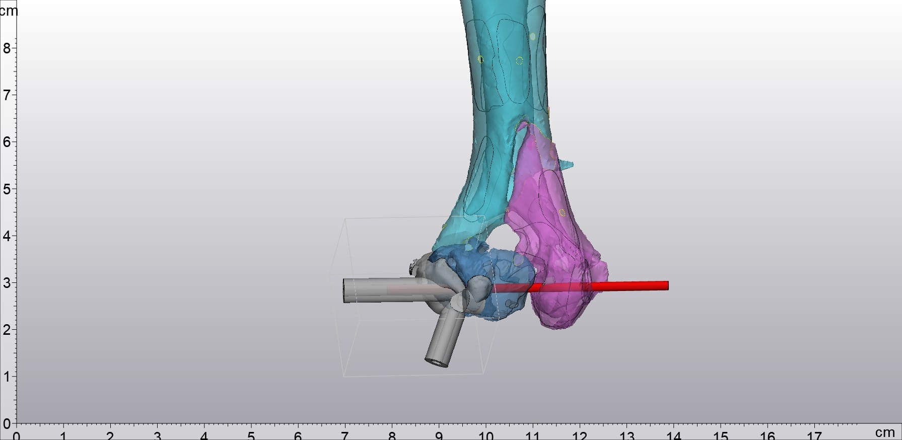
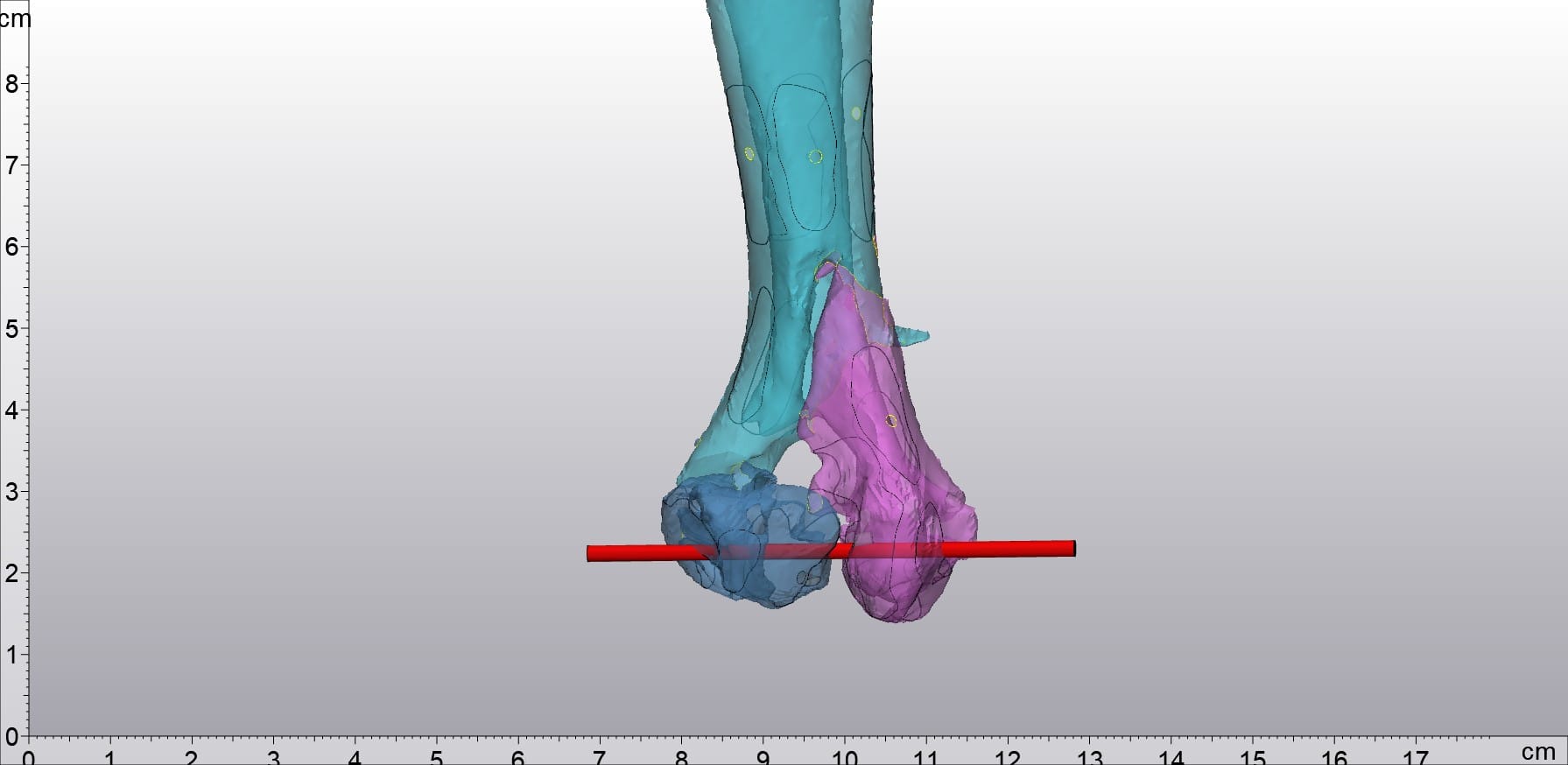
The image above shows a model of a bone pin integrated into the bone, created by Dr. Sebastian.
The dual-material printing capability further expands its range of applications. Dr. Sebastian can now print combined models that display both bone and implants—accurately representing the post-operative state. He can drive bone pins directly into these combined models, simulating the optimal surgical approach before the patient is on the operating table.
| Results and Outlook
The Bambu Lab H2D has had an immediate and measurable impact on Dr. Sebastian's workflow. Anatomically faithful models make surgical rehearsal more straightforward and effective. For most animal bone cases, models can now be printed in one go—eliminating assembly steps, reducing post-processing time, and allowing Dr. Sebastian to focus on surgical preparation rather than model creation.
Multi-material printing further expands possibilities, enabling more complex and realistic simulations that directly translate into better-prepared surgeries.
Future Outlook
Dr. Sebastian plans to fully integrate 3D printing technology into his clinic and establish a comprehensive library of anatomical and pathological models for common and rare conditions, to be used for team training, practical drills, and intern education. His ambitious goal is to build an efficient, sustainable ecosystem for additive manufacturing applications in veterinary medicine.
The H2D enables Dr. Sebastian to work faster, produce more precise models, and be better prepared for complex surgeries—all these improvements ultimately serve one purpose: to deliver better treatment outcomes for his animal patients.
Learn more about Bambu Lab products 👈
- Contact Us -
3DMart offers more than just 3D printing; we provide three major contract services: "3D Printing Service", "3D Scanning Service", and "Spatial 3D Scanning Service"!!
Follow our fan pages for the latest news:
Facebook | Instagram | Threads